BRIEF HISTORY
This is a case of AA, 44 year-old, female, non-hypertensive, non-diabetic, with history of right nephrectomy due to paraganglioma, came in with a 3-month history of throbbing headache and a CT scan that showed tumor mass at the right temporoparietal region. Physical and neurologic examinations were normal. Brain MRI with contrast (Fig. 1) was done showing mass at the right temporal convexity region. Craniectomy with excision of tumor mass was scheduled with preoperative diagnosis of astrocytoma vs oligodendrohygroma.
 Figure 1. Brain MRI with contrast. There is a 5x4 cm dural basal heterogenous enhancing mass at the right temporal convexity region with mass effect and temporoparietal vasogenic edema. There is a midline shift to left, compression of right lateral ventricle and dilatation of the left lateral ventricle. Radiologic impression is right temporal convexity mass, consider meningioma with subfalcine herniation.
OPERATIVE PROCEDURE
Craniectomy of right fronto-temporo-parietal and excision of temporoparietal mass was performed for this patient under general anesthesia.
Figure 2. Curvolinear incision at the right frontotemporo-parietal area was made exposing the skull.


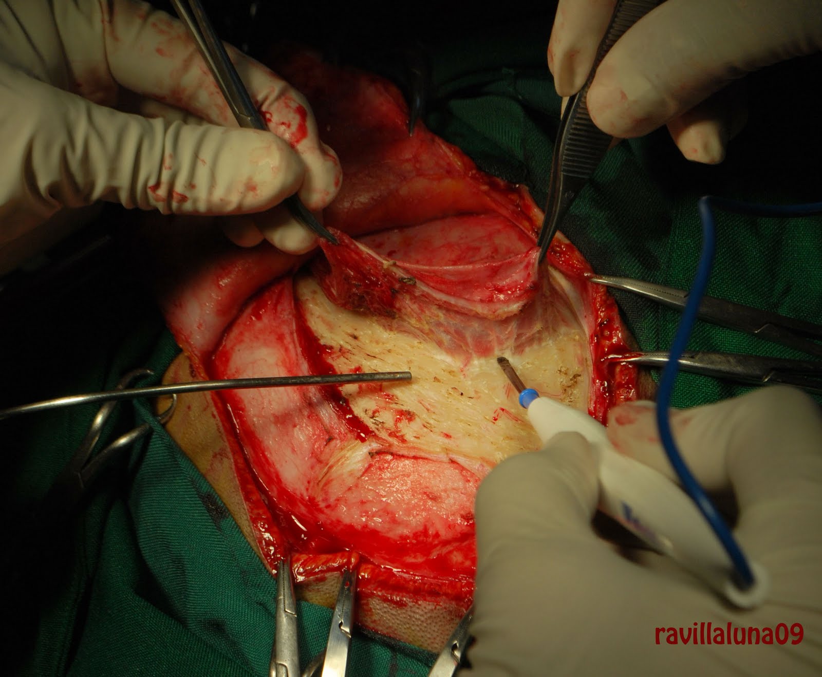
Figure 3. Burr holes were made and craniectomy of right frontotemporo-parietal.
Figure 2. Curvolinear incision at the right frontotemporo-parietal area was made exposing the skull.


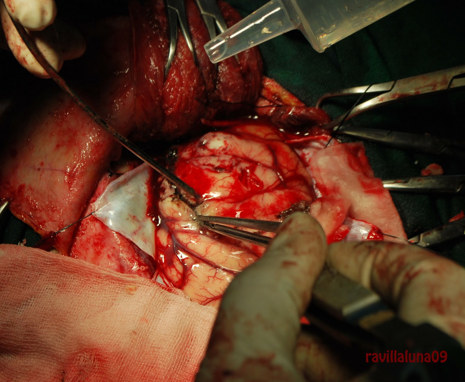


The post operative diagnosis is to consider meningioma. Still awaiting histopathologic result.
What could be the final diagnosis for this case?
.jpg)